
In vivo and ex-vivo 3D high-resolution imaging for inner ear visualization, metabolomics, inflammation, and drug biodistribution.
- ex-vivo 3D imaging
-
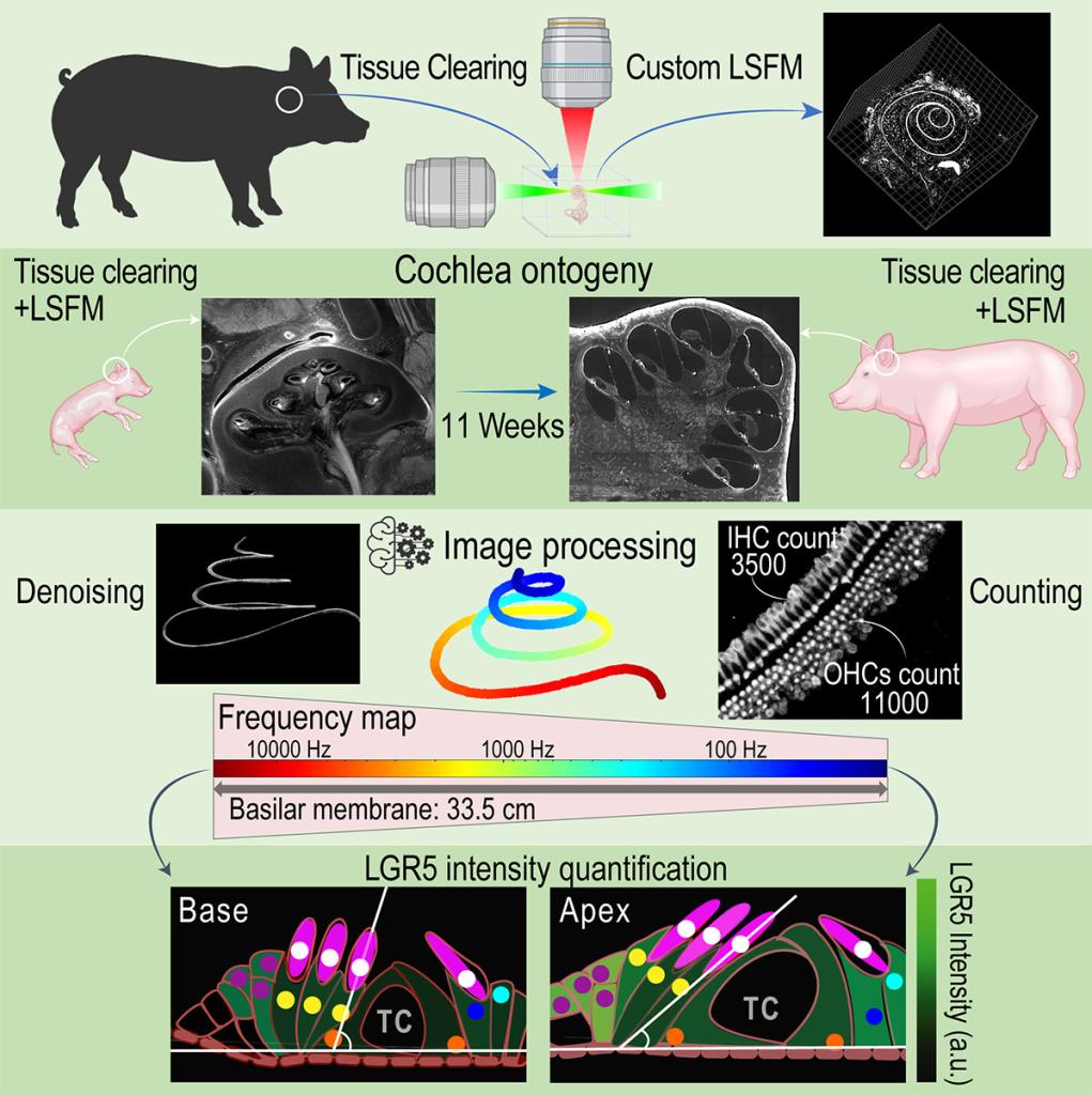
Tissue clearing is a technique that makes biological tissues transparent by removing lipids, pigments, and calcium which are the main cause of opacity. This process allows to visualize 3D structures within tissues without slicing them, preserving spatial relationships. Various methods, like CLARITY, CUBIC, and BoneClear use chemicals to clear tissues and match their refractive index to surrounding solutions for transparency. Cleared tissues are then imaged with advanced microscopy, such as lightsheet microscopy, which is particularly useful in studying complex cellular structures.
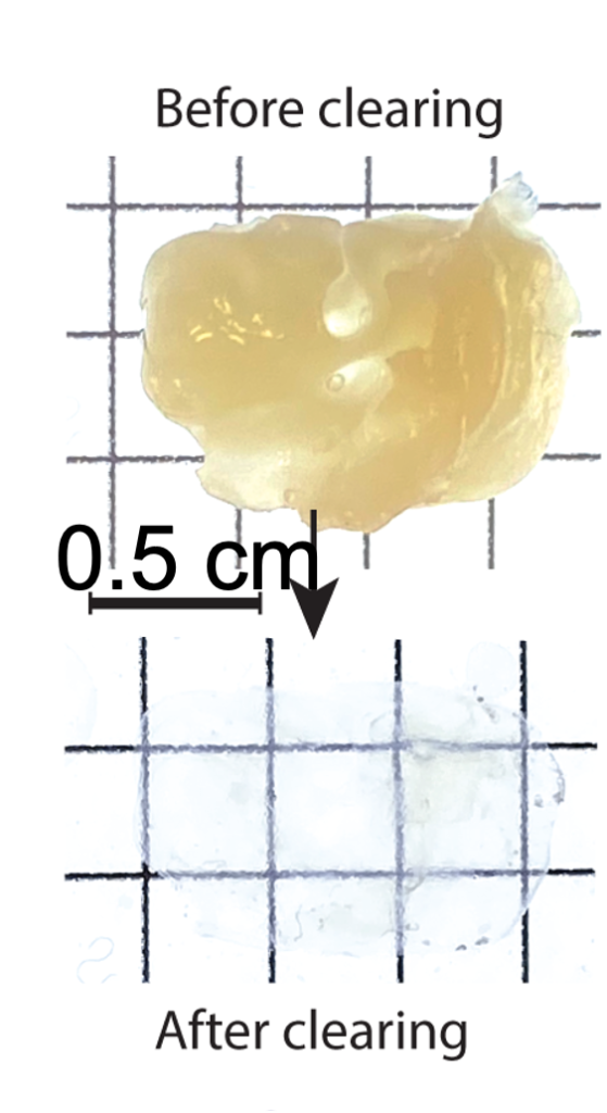
Using tissue clearing technique specifically those designed for bone clearing, we can image the entire inner ear of small and large animal models and humans.
- in-vivo imaging
-
Imaging the inner ear in vivo is difficult because it's residing deep within a dense bone, contains tiny, delicate structures, and is highly sensitive to movement. Additionally, light struggles to penetrate surrounding tissues, making high-resolution, non-invasive imaging nearly impossible.
We can develop biomarkers to improve the resolution of current imaging modalities and detect:
- inflammation
- metabolic state